
Fig. 9.
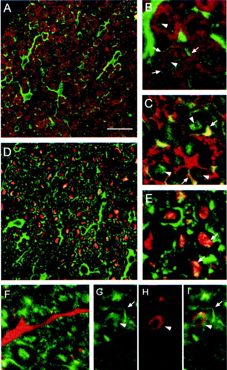
Localization of Nogo-A in CNS white matter by confocal microscopic analysis. A, No colocalization of Nogo-A (shown by AS 472 in green) was found with MBP (red), which is a major constituent of compact myelin. Nogo-A was expressed in oligodendrocyte cell bodies, their processes, and the inner loop (B, C,arrowheads) and outer loop (B, C,arrows) of the myelin sheath. The fixation protocol required to demonstrate MBP expression lowered the Nogo-A signal in the inner loop of the myelin membrane, whereas the different protocol used for MAG and MOG immunohistochemistry showed strong expression of Nogo-A in the inner and outer loops of the myelin sheath.E, Nogo-A (AS 472, green) is expressed in the outer loop (arrows) of the myelin sheath and was found to colocalize there with MOG (red).G, A strong Nogo-A signal (AS 472, green) was also found in the inner loop (arrowheads), where MAG (H, red) is expressed. C, D, Some Nogo-A (AS 472, green) was also present in axons, as was demonstrated by colocalization (yellow) of AS 472 with an antibody against neurofilament (red). F, Nogo-A (AS 472,green) was not present in astrocyte processes stained by an antibody against GFAP (red). Scale bar: A, C, 85 μm; B, D–I, 45 μm.