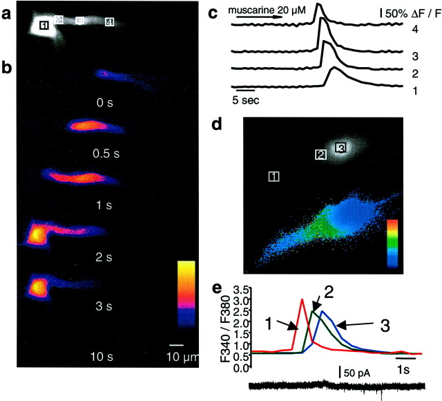
Fig. 1.
Muscarinic stimulation evokes calcium waves in hippocampal pyramidal neurons. A fluorescent image of a cell loaded with 50 μm Oregon Green BAPTA-1 is shown ina. The boxes indicate the regions in which fluorescence measurements were taken. The nucleus can be seen as a bright area in the soma (area 1). b, Selected pseudocolor frames are baseline-subtracted images (ΔF) of the cell shown in aduring bath application of 20 μm muscarine. The first detectable increase in fluorescence is labeled as time 0, and subsequent frames at the indicated times are shown below.c, Rises in calcium, plotted as ΔF/F, measured over the regions indicated in a are shown over time. The calcium transient had the fastest time course in the dendrite and became slower as it propagated to the soma and nucleus. d, Different cell loaded with the ratiometric indicator fura-2 AM (100 μm). The panel above shows image recorded using the isobestic 360 nm excitation wavelength. Theboxes indicate the regions in which fluorescence measurements were made. The selected pseudocolor frame below shows the calcium wave propagating into the soma before entry into the nucleus.e, Rises in calcium plotted as the ratioF340/F360over time for the three regions (dendrite, soma, and nucleus) indicated in the frames above. Note that [Ca2+]iis similar in the soma and nuclease, and there is a clear delay between the calcium rises in the extranuclear soma surrounding the nuclear region and the calcium rise in the nucleus itself.

