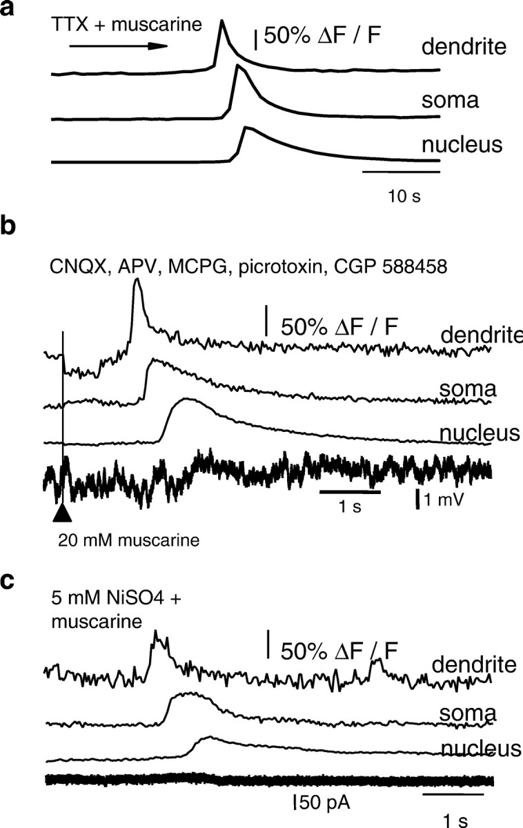
Fig. 2.

Calcium waves are not the result of enhanced network activity. a, Rises in calcium, plotted as ΔF/F, to bath application of muscarine in the presence of TTX (500 nm) are shown ina. b, Focal application of muscarine onto the soma and proximal dendrite readily evoked calcium waves, even when glutamatergic and GABAergic synaptic activity was blocked by bath application of APV, CNQX, MCPG, picrotoxin, and CGP 588458. Rises in calcium are plotted as ΔF/F and are shown at the top. This neuron was recorded in current-clamp mode. The simultaneously recorded voltage is shown below. The timing of the Picospritzer activation is indicated by thearrowhead and the vertical line. Note the delay between application of muscarine and the onset of the calcium wave. c, Calcium waves do not depend on extracellular calcium because a calcium wave could be evoked in 5 mmNiSO4. Rises in calcium are plotted as ΔF/F and are shown for the dendrite, soma, and nucleus, along with the simultaneously recorded whole-cell current.
