Fig. 4.
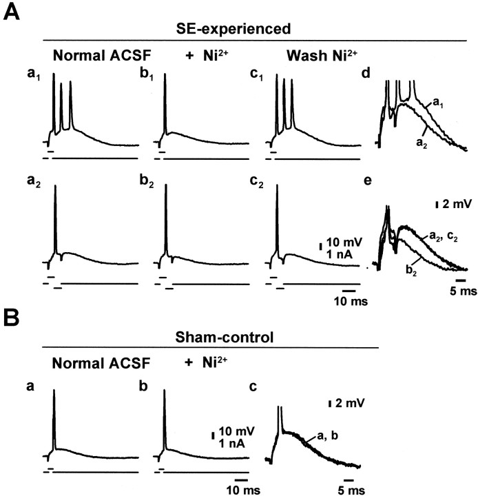
Low concentrations of Ni2+suppress burst firing by reducing the spike ADP. A, Intrinsically bursting CA1 pyramidal neuron from an SE-experienced animal in normal ACSF (a1), after block of the burst discharge by perfusion of 50 μm Ni2+(b1), and after washout (c1). The slow depolarization underlying the burst was unmasked by delivering a brief (4 msec) hyperpolarizing current pulse immediately after the first spike (a2–c2). The time course of the ADP was similar ina1 anda2 (for comparison at larger magnification, see d). Adding 50 μmNi2+ to the ACSF reversibly suppressed the ADP (b2,c2; for comparison at larger magnification, see e). B, Regular firing CA1 pyramidal cell in a sham-control animal before (a) and after (b) application of 100 μm Ni2+. The ADP was not affected by Ni2+ (for comparison at larger magnification, see c).

