
Fig. 4.
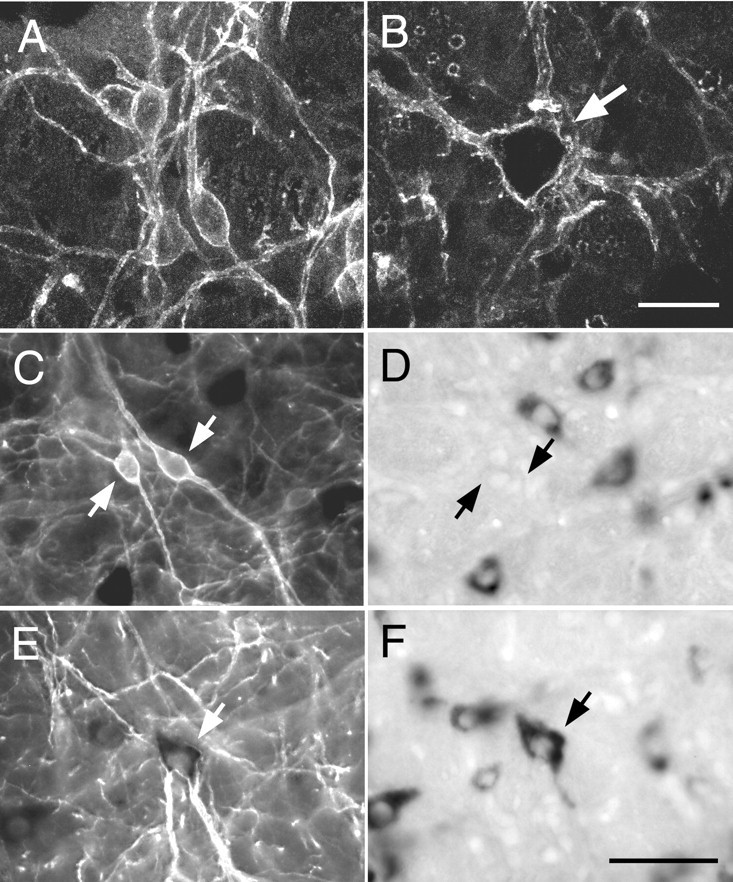
Cell size and presence of PPE mRNA define two types of NK1R-ir neurons in the VRG. A,B, Confocal images of a cluster of small NK1R-ir neurons within the pre-BötC (A) and of a large isolated NK1R-ir neuron of the rVRG (B,arrow). The inside of the cell shown in Bis especially dark because Cy3 fluorescence is quenched by dense VGLUT2 mRNA reaction product. C, D, Light microscopic images of a cluster of small neurons of the pre-BötC under fluorescence (C) and bright-field illumination (D). The NK1R-ir cells (C, arrows) are devoid of PPE mRNA (D) but are surrounded by neurons lacking NK1R immunoreactivity that express high levels of PPE mRNA.E, F, Light microscopic images of a large NK1R-ir neuron of the rVRG under fluorescence (E) and bright-field illumination (F). The dendrites of this cell display intense NK1R immunofluorescence (E), whereas in the cell body, NK1R immunofluorescence is quenched by the very high level of PPE mRNA reaction product (F). Scale bars (shown inB): A, B, 20 μm; (shown in F): C–F, 50 μm.