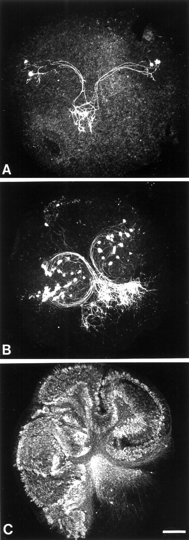
Fig. 1.

Definition of three separate groups of Purkinje cell survival in organotypic culture. Photomicrographs of P3 mouse cerebellar slices, nontreated (control; A) and treated with IGF-I (B) or the PKC inhibitor Gö6976 (C) are shown. These slices were maintained for 5 d in vitro and immunostained with anti-CaBP antibodies to label Purkinje cells. A, Very few Purkinje cells are present, without groups of >20 Purkinje cells. This slice was included in group I. B, In this slice, there is at least one cluster of >20 Purkinje cells (group II). C, The slice contains a cluster of >50 Purkinje cells and was included in group III (see Materials and Methods). Scale bar, 250 μm.
