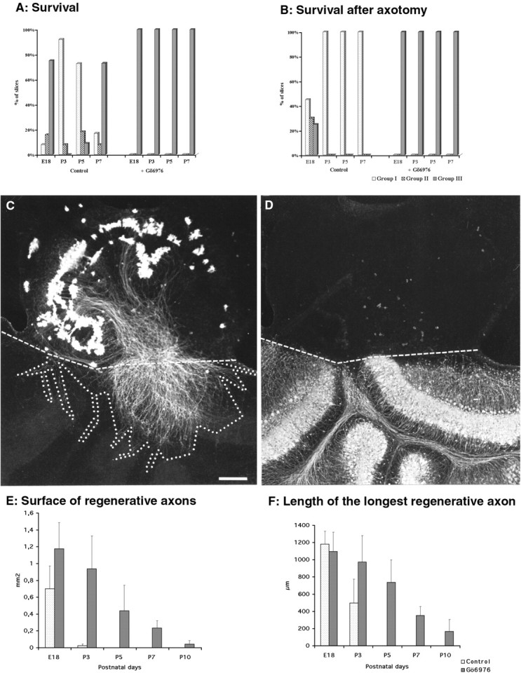
Fig. 5.

Purkinje cell survival and regeneration after axotomy in the presence or absence of Gö6976. A, B, Quantitative evaluation of Purkinje cell survival in organotypic cultures (A) and after axotomy (B). The three groups of slices (I–III) were defined according to the number of Purkinje cells and immunostained with anti-CaBP antibodies as described in Materials and Methods and Figure 1. Interestingly, although without axotomy (A) most of the E18 and P7 slices were in group III (with high Purkinje cells survival), after axotomy (B) they were in group I (with low Purkinje cell survival). However, with or without axotomy, high Purkinje cell survival is always improved by the treatment of the slices with 2 μm Gö6976. C, D, Coculture of the dorsal region of a wild-type P0 mouse cerebellar slice with the dorsal region of split P10 cerebellar slices taken from a CaBP-null mutant. The regenerative axons are CaBP-immunostained (C), whereas the P10 Purkinje cells in the CaBP-null mutant are parvalbumin-immunostained (D). The double labeling permits evaluation without ambiguity of the limits between the two cultures (dashed lines). The area delineated by the dotted linein C represents an example of how we have drawn the surface occupied by the regenerative axons. Scale bar, 200 μm.E, Means of the surface covered by regenerative axons (an example of such a surface is presented in C) with or without Gö6976 at different ages. F, Mean of the lengths of the three longest regenerative axons per slice.
