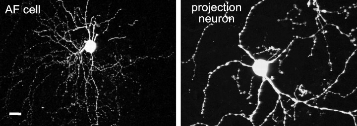
Fig. 14.
The morphological similarity between AF cells and the DLM-projecting cells of area X. The AF cell was recorded via the whole-cell method from a zebra finch brain slice and filled with biocytin; the area X projection neuron was labeled by injection of a retrogradely transported tracer (tetramethylrhodamine-conjugated dextran) into the DLM of a different male zebra finch. Both images were acquired with a confocal microscope (the biocytin fill was visualized using Cy2-conjugated streptavidin) and are shown at the same scale. Scale bar, 10 μm.

