Fig. 2.
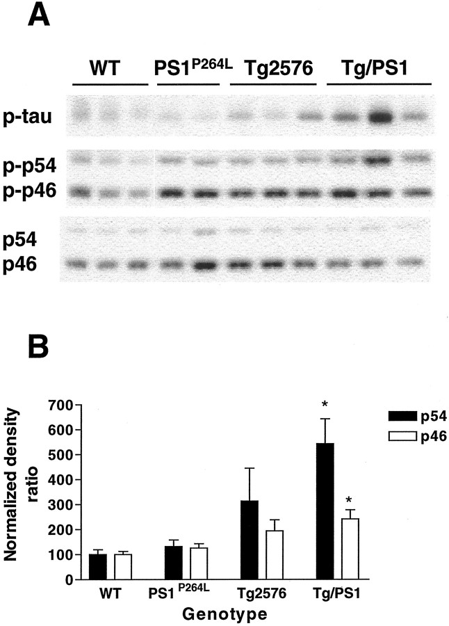
JNK activation is genotype dependent.A, The middle demonstrates cortical samples at 12 months stained as in Figure 1 for detection of activated JNK. Increased phosphorylation of p54 JNK in the Tg2576/PS1P264L cortex compared with wild-type, Tg2576, or PS1P264L cortex. Thebottom depicts total JNK as in Figure 1. Thetop shows the same samples stained for a third time for phosphorylated tau at ser202. Note the increase in phospho-tau in the Tg2576/PS1 cortex with the greatest amyloid burden and an intermediate degree of phospho-tau in Tg2576. B depicts a significant increase at p ≤ 0.05 (asterisks) in the active fraction of both p54 and p46 JNK in the Tg2576/PS1P264L cortex compared with wild type. Three samples per group were assayed in triplicate on immunoblot.

