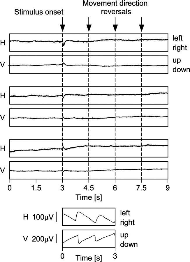
Fig. 3.
Absence of eye movements during recording of neuronal activity for correlation measurements. The topof the figure shows three pairs of horizontal (H) and vertical (V) eye movement recordings. Thetopmost pair was recorded under monocular stimulation of the dominant eye, the middle pair under dichoptic stimulation, and the bottom-most pair under monocular stimulation of the nondominant eye. Stimulus onset is at 3 sec after the start of the recording, and movement direction of the stimuli reverses every 1.5 sec, as indicated by the arrows andbroken lines. Evidently, the traces do not reveal OKN or any other pursuit movements. The short-latency deflections after stimulus onset are with all likelihood caused by light-induced potential changes in the retina. The bottom of the figure displays, at the same scale, recordings of OKN that were obtained from the same animal in the same recording session during prolonged stimulation without movement direction reversals (compare with Fig. 1). It should be noted that the two OKN recordings were not made simultaneously. For the induction of horizontal OKN (top trace), the dominant (left) eye was stimulated monocularly in temporonasal direction, and for the vertical OKN (bottom trace), the dominant eye was stimulated monocularly in the upward direction.

