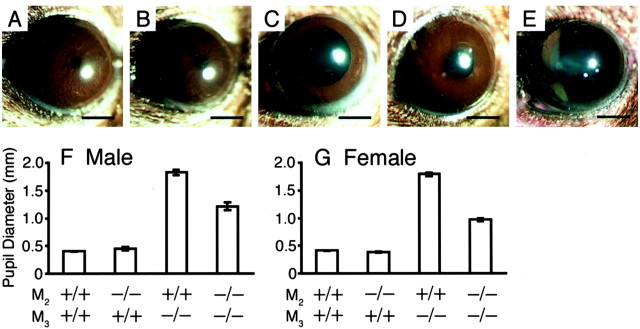
Fig. 5.
Appearance of pupils of the wild-type (A), M2−/− (B), M3−/− (C), and M2−/−M3−/− (D) mice. The pupils of the M2−/−M3−/− mice (D) are smaller than those of the M3−/− mice (C). Full mydriasis was achieved in the M2−/−M3−/− mouse after instillation of atropine solution (E). The pupils of the M2−/− mice (B) are indistinguishable from those of the wild-type mice (A). Scale bars, 1 mm. F,G, Pupil diameter (mean ± SEM;n = 4–30) of the mutant mice, measured in a bright room (∼1000 lux). The M2−/−M3−/− mice have smaller pupils than the M3−/− mice in both sexes.

