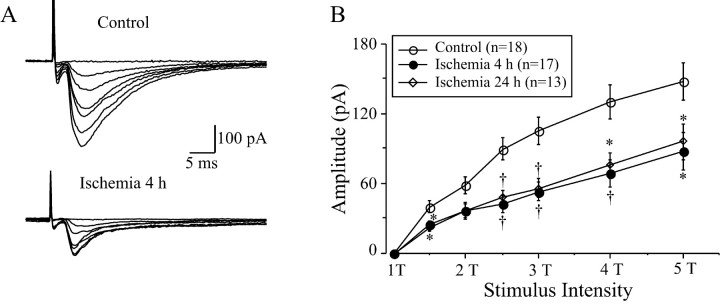
Fig. 3.
Changes in EPSCs in LA neurons before and after ischemia. A, Representative traces recorded from LA neurons before and at 4 hr after ischemia. The amplitude of evoked EPSC increased with increasing stimulus intensities (0–5 times threshold intensities, 0–5T), but the amplitude of EPSCs at 4 hr after ischemia was significantly smaller than that of control ones. The traces are the average of six consecutive recordings. B, The input–out relationship of evoked EPSCs before and at different intervals after ischemia. The amplitude of EPSCs was dramatically decreased at 4 and 24 hr after ischemia at all stimulus intensities (1.5–5T). *p < 0.05;†p < 0.01.

