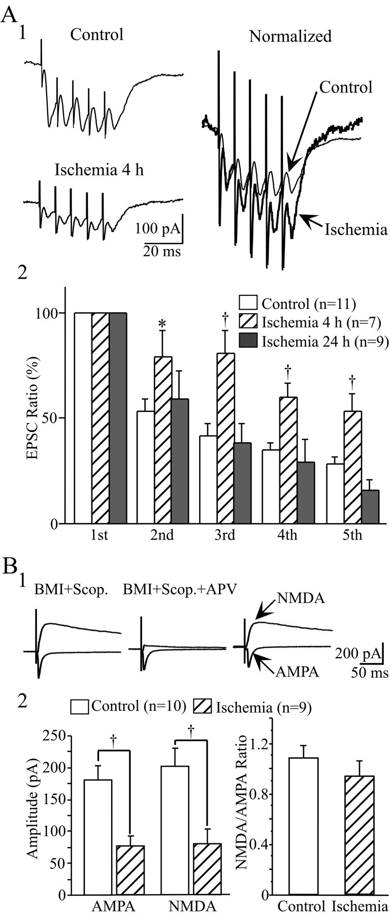
Fig. 6.

Comparison of EPSCs elicited by a train of high-frequency (100 Hz) stimulation and changes in NMDAR/AMPAR-mediated responses before and after ischemia. A1, Representative traces showing the synaptic responses induced by a train of five stimuli before ischemia and at 4 hr after reperfusion. The two traces were normalized to the same amplitude of the first EPSCs to show the difference in the fifth/first EPSC ratio between these traces (right). All traces are the average of six consecutive recordings. A2, Pooled data showing the changes in EPSCs during train stimuli. All amplitudes were normalized to the first EPSCs at each recording. The fifth/first EPSC ratio was significantly higher at 4 hr after ischemia compared with controls and at 24 hr after ischemia, suggesting that the releasing probability of LA neurons was transiently decreased shortly after ischemia. B, Parallel change in NMDAR-and AMPAR-mediated responses before and at 4 hr after ischemia. B1, left, Evoked EPSC in the presence of 20 μm BMI and 2 μmscopolamine (Scop.), at the holding potentials of −80 mV and +60 mV, respectively. Middle, Evoked AMPAR-mediated responses in the presence of BMI, scopolamine, and 50 nmd-APV. Right, top trace, The NMDAR-mediated response was obtained by subtracting the left two top traces. B2, left histogram, Parallel decrease in NMDAR- and AMPAR-mediated responses after ischemia. Right histogram, Comparison of NMDAR-mediated current with the AMPAR-mediated current ratio before and after ischemia. No significant difference in NMDA/AMPA ratio was detected between these two conditions. *p < 0.05;†p < 0.01.
