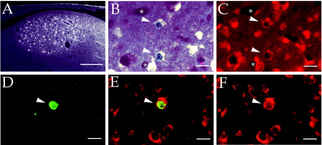
Fig. 2.
A, Low-magnification fluorescence (UV) photomicrograph of the HVC after retrograde labeling with Fluoro-Gold injections into the RA. B, Higher-power magnification of two HVC-RA projection neurons formed in adulthood (arrowheads) viewed with combined bright-field and UV illumination. These neurons have developed silver grains overlying their nucleus and Fluoro-Gold in their cytoplasm. C, Same view as in B, showing all cells counterstained with fluorescent cresyl violet and viewed under combined rhodamine fluorescence and bright-field optics. Arrowheads point to the same 3H-labeled neurons shown in B.Asterisks in B and C label blood vessels cut in cross section. D–F, The same field is shown using different fluorescence filters to reveal a cell double labeled with BrdU (green) and Hu (red; arrowheads). D, BrdU-labeled cell nucleus viewed under FITC fluorescence.E, Same field viewed with dual FITC–rhodamine filter. Cytoplasmic staining with the neuronal marker Hu surrounds the BrdU-labeled cell nucleus. F, Same field viewed under rhodamine fluorescence. Scale bars: A, 100 μm;B–F, 10 μm.

