
Fig. 7.
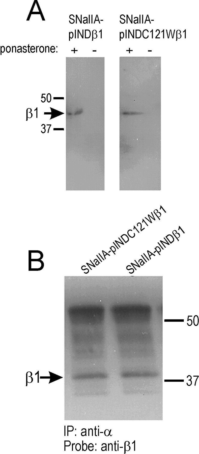
Ecdysone-inducible expression of β1 or C121Wβ1 subunits and association of β1 or C121Wβ1 with rat Nav1.2a α subunits. A, SNaIIA-pIND.β1 or SNaIIA-pIND.C121Wβ1 cells were treated with vehicle (0 ponasterone) or hormone (20 μm ponasterone) for 48 hr in culture, solubilized in 5% SDS, and boiled in SDS-PAGE sample buffer containing 5% β-mercaptoethanol. Samples were separated by 10% acrylamide SDS-PAGE and transferred to nitrocellulose. Western blots were probed with anti-β1EX antibody (1:500 dilution) and then with horseradish peroxidase-conjugated goat anti-rabbit antibody (1:100,000). Immunoreactive bands were visualized with Westdura chemiluminescent substrate. Arrow indicates position of β1 immunoreactive band. B, Equal aliquots of SNaIIA-pIND.β1 or SNaIIA-pIND.C121Wβ1 cells were treated with 20 μm ponasterone for 48 hr in culture, and then equal aliquots of cells were immunoprecipitated with anti-Nav1.2a antibody as described in Materials and Methods. The samples were then separated by SDS-PAGE, transferred to nitrocellulose, and probed with anti-β1EX antibody (1:500), followed by horseradish peroxidase-conjugated goat anti-rabbit antibody (1:100,000). The blot was detected with Westdura chemiluminescent substrate and exposed to ECL Hyperfilm. Arrow indicates migration of immunoreactive β1 subunits.