Fig. 1.

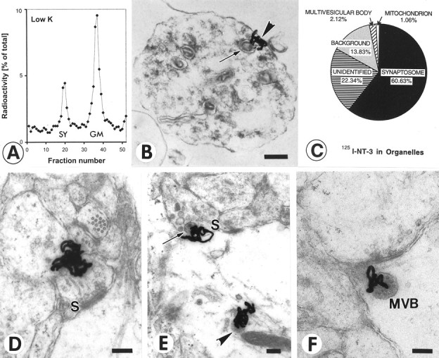
Quantification of radiolabeled NT-3 in retinotectal terminals after anterograde axonal transport.A, Quantification of 125I-NT-3 in the synaptosomal peak after centrifugation on ficoll-sucrose gradients and fractionation. The synaptosomal peak (SY) and the general membrane fraction (GM) are indicated. B, Synaptosome shows a silver grain (arrowhead) next to a large dense core vesicle (arrow). C, Quantification of silver grains over synaptosomes shows majority of grains over synaptosomes. Silver grains (n = 94) were randomly analyzed. D, Synapse before release of NT-3. Cluster of silver grains over a presynaptic terminal in layer f of the stratum griseum et fibrosum superficiale (SGFSf) of the optic tectum after anterograde transport. Note the synaptic profile (S). E, Synapse in the process of releasing NT-3. Silver grains over a presynaptic profile (S) containing a large dense core vesicle (arrow) and over a postsynaptic organelle reminiscent of a multivesicular body (arrowhead). F, Synapse after release of NT-3. Silver grain on the postsynaptic side of the synapse (S) overlying a multivesicular body (MVB). Scale bars, 200 nm.
