
Fig. 6.
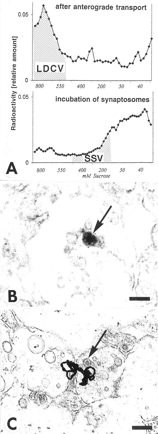
Association of NT-3 with large dense core vesicles (LDCV) rather than small synaptic vesicles (SSV) in retinotectal terminals. A, Vesicle purification of tecta containing anterogradely transported125I-NT-3 shows higher levels of radioactivity in sedimentation fractions known to contain LDCVs in chick embryo brains (Han and Fischbach, 1999), but not in fractions known to contain SSVs (top panel). When homogenized tecta were incubated with125I-NT-3 (bottom panel), radioactivity was seen in sedimentation fractions known to accumulate SSVs (Han and Fischbach, 1999). B, Section through a retinotectal terminal from a 17-d-old chick embryo immunolabeled with an antibody to NT-3, processed with horseradish-peroxidase, and visualized with diaminobenzidine. Note reaction product in a large dense core vesicle (arrow). Scale bar, 100 nm. C, Section through the stratum opticum (SO) of the optic tectum after injection of125I-NT-3 in the eye. Note a cluster of silver grains in the vicinity of a large dense core vesicle (arrow) in sectioned retinotectal fibers. Scale bar, 500 nm.