
Fig. 1.
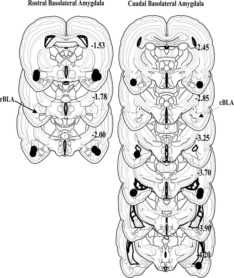
Schematic representing coronal sections of the rat brain and cannula placements within the rostral (left) and caudal (right) regions of the basolateral amygdala.Circles indicate the location and diffusion of lidocaine and are drawn to scale. All drawings are based on the atlas of Swanson (1992) with the anterior–posterior reference measured from bregma. Each placement is shown at its midpoint along the anterior–posterior plane.