Fig. 7.
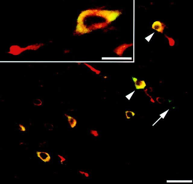
Neurons sustaining TAI demonstrate a somatic increase in eIF2α(P). Double labeling with antibodies to β-APP (red) and eIF2α(P) (green) reveal a striking increase in somatic eIF2α(P) in neurons with axonal injury (two examples are indicated with arrowheads). However, isolated neurons exhibiting no evidence of TAI or somatic increase in APP were also found to demonstrate a somatic increase in eIF2α(P) immunofluorescence (arrow). Scale bar, 50 μm. Inset, Higher magnification of neuronal soma with TAI. Increased immunofluorescence for eIF2α(P) can be seen to colocalize to the cytoplasm of the neuronal cell body. Scale bar, 20 μm.

