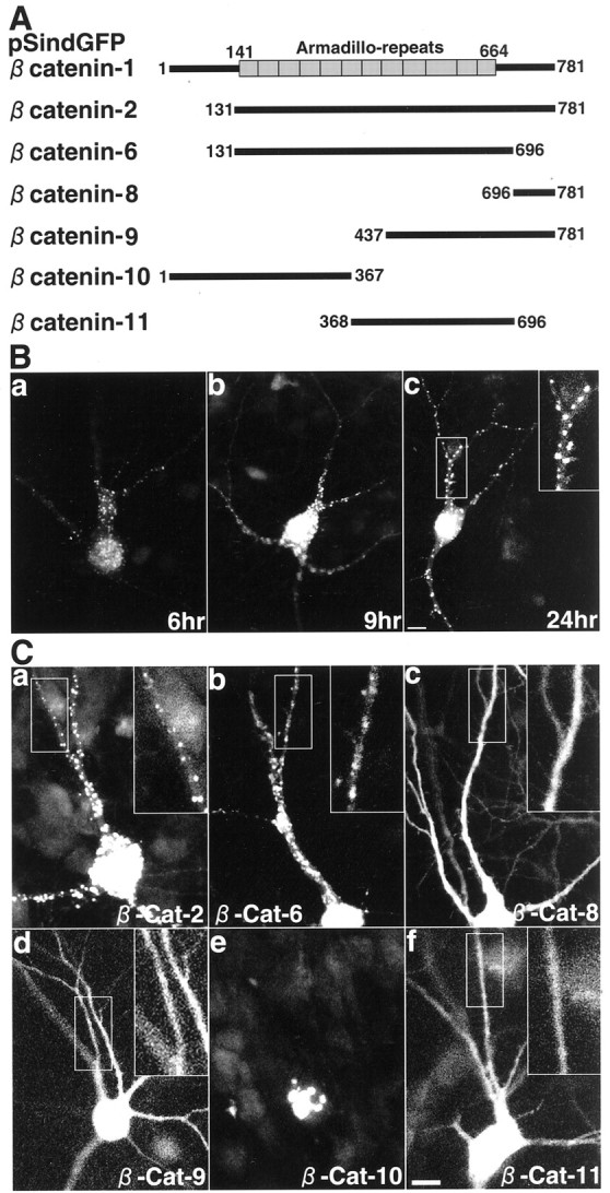
Fig. 6.

Various GFP–β-catenin proteins expressed in hippocampal slice neurons. A, Schematic of various GFP-tagged constructs of β-catenin. Boxes, Armadillo repeats. B, Temporal profile of the expression of GFP–β-catenin-1 in hippocampal slice neurons.a, 6 hr after the infection; b, 9 hr after the infection; c, 24 hr after the infection.Inset, Demarcated area at higher magnification. Scale bar, 10 μm. C, Various GFP–β-catenin proteins in hippocampal slice neurons. Insets, Demarcated areas at higher magnification. a, GFP–β-catenin-2;b, GFP–β-catenin-6; c, GFP–β-catenin-8; d, GFP–β-catenin-9;e, GFP–β-catenin-10; f, GFP–β-catenin-11. Scale bar, 10 μm.
