
Fig. 2.
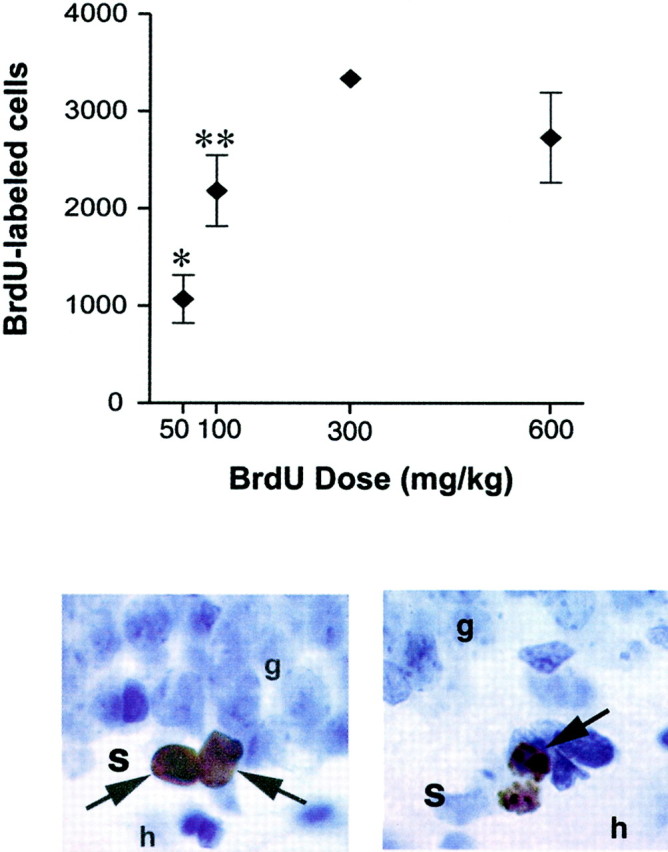
Methodological considerations related to BrdU labeling in adults. Top panel, A short pulse of BrdU labels in a dose-dependent manner, raising the possibility that changes in the number of BrdU-labeled cells could result from differences in BrdU availability. The graph shows the total number of BrdU-labeled cells in the dentate gyrus of adult rats after a single injection of BrdU 30 min earlier. *Indicates significant difference from all other groups, p < 0.05. **Indicates significant difference from 300 mg/kg group, p < 0.05. Adapted from Cameron and McKay (2001) with permission. Bottom panel,Left, BrdU-labeled progenitor cells in the subgranular zone (s) of the dentate gyrus. BrdU is an exogenous marker of proliferating cells.Right, Dividing cell undergoing cytokinesis labeled with phosphorylated histone H3 in the subgranular zone. p histone H3 is an endogenous marker of cells in M phase (Hendzel et al., 1997). Endogenous markers can be used to verify that changes in the number of BrdU-labeled cells are attributable to differences in the number of dividing cells, as opposed to difference in BrdU availability.g, Granule cell layer; h, hilus.