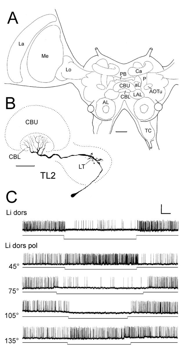
Fig. 1.

Responses of a TL2 tangential neuron of the lower division of the central body to polarized light. A, Frontal diagram of the locust brain, indicating the position and subdivisions of the central complex in relation to other neuropil structures. AL, Antennal lobe; aL, α-lobe of the mushroom body; AOTu, anterior optic tubercle; Ca, calyces of the mushroom body;CBL, CBU, lower and upper division of the central body; La, lamina; LAL, lateral accessory lobe; Lo, lobula; Me, medulla;P, pedunculus of the mushroom body; PB, protocerebral bridge; TC, tritocerebrum. Scale bar, 200 μm. B, C, Frontal reconstruction (B) and intracellular recording (C) of a Lucifer yellow-injected TL2 neuron of the central body. The neuron has its cell body in the inferior median protocerebrum. It innervates the lateral triangle (LT) of the lateral accessory lobe and layer 2 of the lower division of the central body (CBL). Scale bar, 100 μm. C, The neuron is tonically inhibited by dorsal unpolarized light (Li dors). It shows tonic excitation or inhibition to dorsal polarized light (Li dors pol) depending on e-vector orientation. Calibration: 10 mV, 1 sec.
