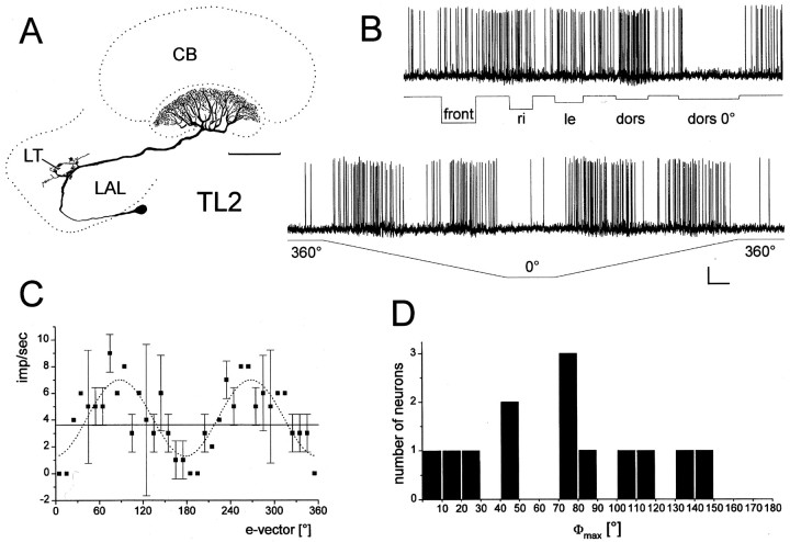
Fig. 2.
Responses of TL2 neurons to unpolarized and polarized light. A–C, Frontal reconstruction (A), intracellular recording (B), and e-vector response plot of a TL2 neuron, studied in an isolated head preparation.A, The neuron arborizes in the lateral triangle (LT) of the lateral accessory lobe (LAL) and in layer 2 of the lower division of the central body (CB). Scale bar, 100 μm.B, The neuron does not respond to illumination of the animal from frontal (front), right (re), or left (le) but is tonically excited by dorsal unpolarized light (dors). The neuron is completely inhibited by dorsal polarized light withe-vector orientation at 0° (dors 0°). Rotation of the polarizer through 360° results in alternating excitations and inhibitions, independent of turning direction. Calibration: 5 mV, 2 sec. C, e-vector response plot (means ± SD; n = 2, one clockwise and one counterclockwise rotation). Solid lineindicates background activity. Fitting the data to a sin2-function (dotted line) reveals a Φmax of 88.4°. D, Distribution of Φmax from 13 recorded TL2 neurons.

