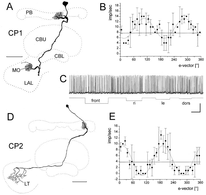
Fig. 7.
CP1 and CP2 neurons are polarization-sensitive.A–C, Frontal reconstruction (A),e-vector response plot (B), and responses to unpolarized light stimuli of a CP1 neuron.A, The CP1 neuron has its soma in the pars intercerebralis. It innervates the innermost column L8 in the left hemisphere of the protocerebral bridge (PB) and has arborizations throughout the median olive (MO) of the lateral accessory lobe (LAL). B,e-vector-response plot (means ± SD;n = 2). Solid line, Background activity. Sin2-fitting (stippled line) reveals a Φmax of 106.2°.C, The neuron shows no response to unpolarized light stimuli. Calibration: 20 mV, 2 sec. D, E, Reconstruction (D) and e-vector response plot (E) of a CP2 neuron.D, The neuron innervates column L4 in the left hemisphere of the protocerebral bridge and has arborizations throughout the lateral triangle of the lateral accessory lobe (LT). E, e-vector response plot (means ± SD; n = 2).Solid line, Background activity. Sin2-fitting (stippled line) reveals a Φmax of 13.0°.

