Fig. 1.
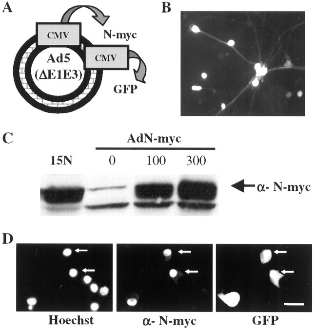
Characterization of a recombinant adenovirus expressing human N-myc. A, Adenovirus construct encoding human N-myc and green fluorescent protein under separate but identical CMV promoters. B, Adenovirus N-myc (AdN)-myc is expressed in sympathetic neurons. Fluorescent micrograph of GFP expression in postmitotic sympathetic neurons 72 hr after infection with 300 MOI of AdN-myc. C, Western blot of equal amounts of protein isolated from LAN-1–15N neuroblastoma cells and from sympathetic neurons infected for 48 hr with 0–300 MOI of N-myc adenovirus and probed with an antibody for N-myc. Note that uninfected sympathetic neurons express N-myc and that LAN-1–15N cells express levels of N-myc similar to sympathetic neurons infected with 100–300 MOI of the N-myc adenovirus. D, Photomicrographs of sympathetic neurons infected with AdN-myc and then analyzed for expression of GFP and immunocytochemically for expression of N-myc (α-N-myc). Cells were also stained with the nuclear dye Hoechst 33258 to identify all of the cells in the field. Note that cells that are GFP positive are also overexpressing N-myc and that the overexpressed N-myc is primarily nuclear. Scale bar, 50 μm.

