
Fig. 2.
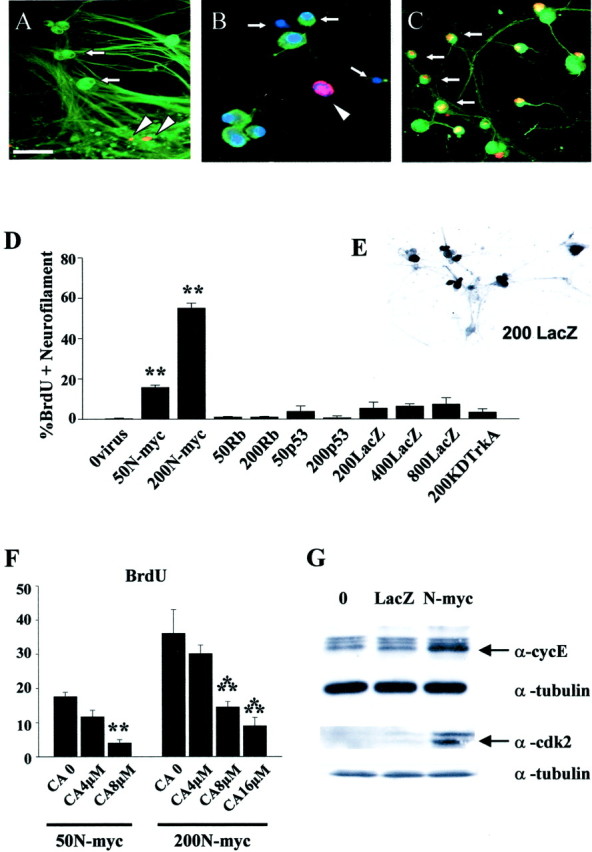
Overexpression of N-myc in postmitotic sympathetic neurons leads to S-phase entry.A–C, Photomicrographs of sympathetic neurons infected with recombinant adenovirus, pulse-labeled with BrdU, and then immunocytochemically analyzed for expression of neurofilament-M (green) and BrdU (red). In some cases, cells were also labeled with Hoechst to label all nuclei (blue). A, AdLacZ-expressing neurons do not incorporate BrdU. Neurons were infected with 200 MOI of a β-galactosidase adenovirus and maintained in 10 ng/ml NGF for 3 d. Note that none of the neurofilament-positive neurons are BrdU positive (arrows) and that the only BrdU-positive cells are neurofilament-negative non-neuronal cells (arrowheads).B, Uninfected neurons that were withdrawn from NGF in the presence of BrdU for 30 hr and then analyzed immunocytochemically. Note that a neurofilament-negative non-neuronal cell with an intact nucleus has incorporated BrdU (arrowhead) but that neurons in different phases of apoptosis are not BrdU positive (arrows). C, AdN-myc-expressing neurons incorporate BrdU. Neurons were infected with 200 MOI of N-myc adenovirus and maintained in the presence of NGF. One day later BrdU was added, and cells were analyzed immunocytochemically 48 hr later. Note that under these conditions, many of the neurofilament-positive neurons are BrdU positive (arrows). Scale bar:A, C, 100 μm; B, 50 μm. D, Quantitation of the percentage of cells positive for both BrdU and neurofilament after infection with various MOIs of recombinant adenovirus in the presence of NGF for 1 d, followed by the addition of BrdU for an additional 48 hr. Neurons were infected with recombinant adenoviruses expressing N-myc, constitutively hypophosphorylated pRb (Rb) (Toma et al., 2000), p53 (Slack et al., 1996), β-galactosidase (LacZ), or a kinase-inactive mutant of TrkA (KDTrkA) (Vaillant et al., 1999). Values derive from counts of at least 300 cells in four or more random microscope fields per condition, and error bars represent the SD of the mean. Similar results were obtained in four independent experiments. **p < 0.01; Student'st test. Note that only N-myc led to BrdU incorporation in sympathetic neurons. E, Staining for β-galactosidase (LacZ) in cultures of sympathetic neurons transduced with 200 MOI of the β-galactosidase adenovirus. Note that most of the sympathetic neurons are positively stained. F, Inhibition of N-myc stimulated cell cycle entry by DNA polymerase inhibition. Percentage of BrdU-positive neurons 48 hr after infection with 50 or 200 MOI AdN-myc, cultured in the presence of BrdU with or without 4, 8, or 16 μmcytosine arabinoside for the final 24 hr. Quantitation was performed as in D. Similar results were obtained in three independent experiments. Concentrations of CA shown here did not increase apoptotic cell morphology over the time of the experiment. **p < 0.05; ***p < 0.001.G, AdN-myc expression increases the levels of the S-phase markers cyclin E and cdk2. Western blot analysis of equal amounts of protein from sympathetic neurons that were uninfected (0) or infected with 200 MOI of adenoviruses expressing either β-galactosidase (LacZ) or N-myc for 2 d. Blots were reprobed for total α-tubulin as a loading control. Similar results were obtained in two independent experiments.