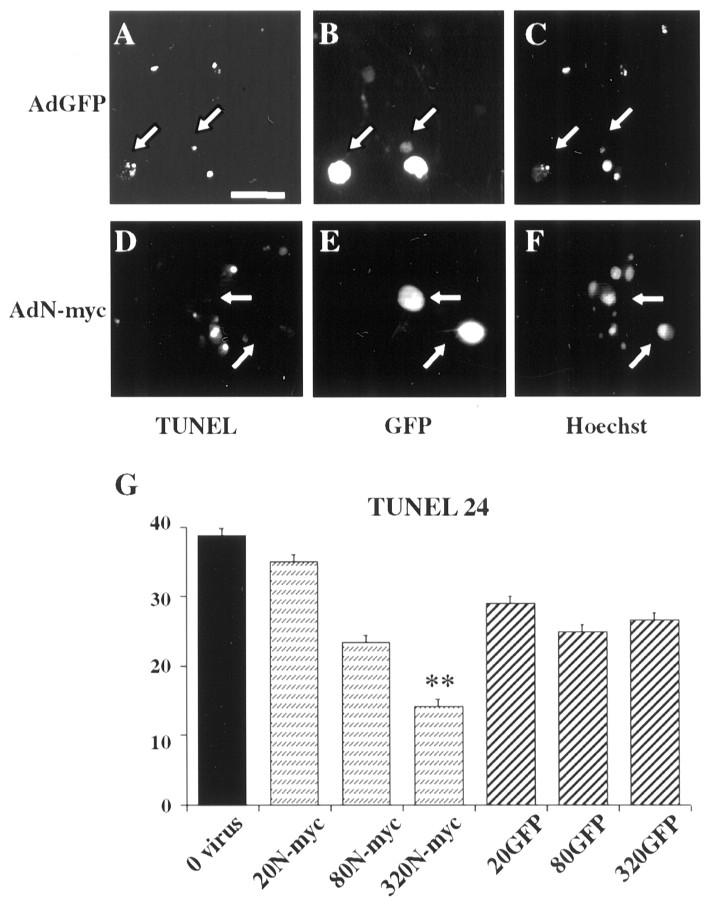
Fig. 4.
N-myc overexpression inhibits sympathetic neuron apoptosis after NGF withdrawal. A–F, TUNEL of sympathetic neurons infected with adenoviruses expressing GFP (AdGFP; A–C) or N-myc (this virus also expresses GFP) (AdN-myc;D–F) and withdrawn from NGF for 48 hr. Photomicrographs show TUNEL-positive nuclei (A, D), GFP-expressing cells (B, E), and Hoechst-positive nuclei (C, F) in the same fields (A–C andD–F). Arrowsindicate the same neurons in each field. Note that neurons infected with the adenovirus expressing only GFP (A–C) are TUNEL positive and display shrunken, apoptotic nuclei, but that those infected with the virus expressing both GFP and N-myc (D–F) are not TUNEL positive. Scale bar, 100 μm. G, Percentage of TUNEL-positive cells 24 hr after NGF withdrawal. Neurons were infected with various MOIs of the N-myc or GFP adenoviruses, and the total number of TUNEL-positive nuclei in three randomly selected fields was counted. The values represent the average of two independent experiments, and error bars represent the SD of the mean. **p < 0.01 in Student's t test comparing apoptotic (TUNEL-positive) cell percentage after infection with AdN-myc versus AdGFP.

