
Fig. 6.
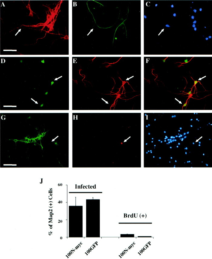
N-myc does not cause BrdU incorporation in postmitotic cortical neurons. A–C, Immunocytochemical analysis of postmitotic cortical neuron cultures. Neurons were cultured as described in Results and double labeled with antibodies for neuron-specific MAP2 (A) and astrocyte-specific GFAP (B). Cells were also stained with Hoechst to show cell nuclei (C). The same field is shown in all three panels, and arrowsindicate the occasional astrocyte present in the cultures. Note that most fields did not contain any GFAP-positive cells.D–F, Cortical neurons were infected with 100 MOI N-myc adenovirus (which also expresses GFP) and then immunostained for MAP2. Visualization of GFP (D) revealed those cells that were transduced with the N-myc adenovirus, whereas immunostaining for MAP2 (E) indicated that the infected cells were neurons. F is the merged image resulting from D and E.Arrows indicate cells that were positive for both GFP and MAP2. G–I, Cortical neurons were infected with 100 MOI N-myc adenovirus and then incubated in the presence of BrdU for 24 hr before double-label immunocytochemical analysis for MAP2 (G) and BrdU (H). The cells were also stained with Hoechst (I). The same field is shown in all three panels, and the arrow indicates a non-MAP2-positive non-neuronal cell that is BrdU positive.J, Left two bars, Quantitation of data showing that N-myc fails to promote S-phase entry in postmitotic cortical neurons. Quantitation is of data similar to those shown inD–F and indicates the percentage of MAP2-positive neurons that were GFP positive after infection with 100 MOI of N-Myc/GFP (100N-myc) or control GFP (100GFP) adenovirus. Right two bars, The percentage of MAP2-positive cells that were BrdU positive after infection with 100 MOI of either the N-myc or GFP adenoviruses. Similar results were obtained in three independent experiments.