
Fig. 1.
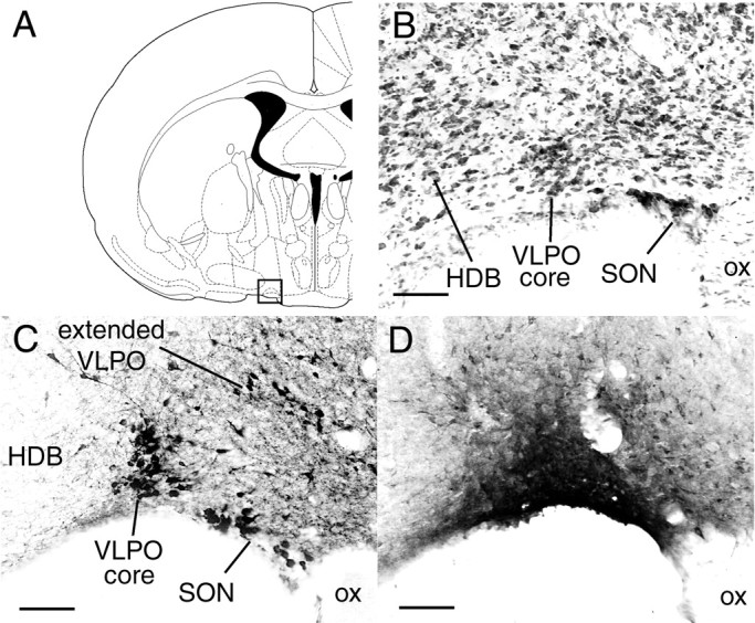
A, Coronal atlas section (Paxinos and Watson, 1997) with square outline showing lateral preoptic region enlarged in B–D.B, Nissl-stained section shows cell-dense VLPO core in relation to adjacent structures such as the horizontal nucleus of the diagonal band (HDB) and supraoptic nucleus (SON). C, Galanin immunostaining shows a dense cluster of immunoreactive neurons in the VLPO core and diffusely distributed neurons in the extended VLPO, which is dorsal and medial to the VLPO core. D, CTB injection into VLPO core (case 40). Scale bars, 100 μm.