
Fig. 4.
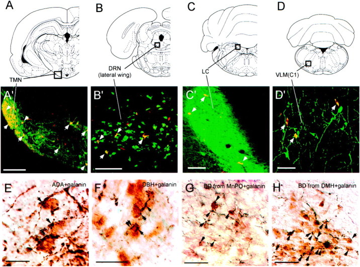
Photomicrographs of double-label immunohistochemistry experiments. A–D(top row) show atlas sections (Paxinos and Watson, 1997) outlining monoaminergic nuclei photographed in panelsA'–D' (middle row). Photographs in A'–D' are from brains with CTB injections into the VLPO core. Redimmunofluorescence indicates CTB-labeled neurons, andgreen immunofluorescence represents the following:A', adenosine-deaminase immunoreactivity (a marker for histaminergic neurons) in the TMN; B', serotonin transporter immunoreactivity in the lateral wing of the dorsal raphe;C', TH immunoreactivity in the locus coeruleus; andD', TH immunoreactivity in the C1 adrenergic cells of the ventrolateral medulla (VLM). Double-labeled cells (yellow) are seen in all four monoaminergic nuclei and are indicated by white arrows.E–H (bottom row) show the VLPO core stained for galanin, shown by brownimmunoreactivity. Black immunoreactive fibers are also seen, representing adenosine-deaminase immunoreactivity (E) or DBH immunoreactivity (F). Apparent appositions of presumably monoaminergic fibers onto galanin-immunoreactive neurons are seen in both cases and indicated by arrows. G andH show black anterogradely labeled fibers arising from biotinylated dextran injections into the MnPO (G) or DMH (H). Apparent appositions of anterogradely labeled fibers onto galanin-immunoreactive VLPO neurons are indicated byarrows. Scale bars: A, 200 μm;B–D, 100 μm; E,F, 20 μm; G, H, 50 μm.