
Fig. 6.
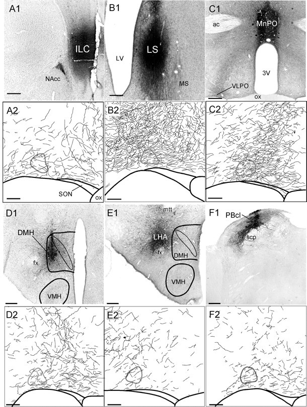
Anterograde tracer injections into infralimbic cortex (A), lateral septal nucleus (B), median preoptic nucleus (C), dorsomedial hypothalamic nucleus (D), lateral hypothalamic area (E), lateral parabrachial nucleus (F). A1–F1, Photomicrographs of BD injection sites. Scale bars, 250 μm.A2–F2, Camera lucida drawings of anterogradely labeled fibers in the VLPO. Scale bars, 100 μm. The VLPO core, identified by Nissl counterstain, is outlinedin gray.