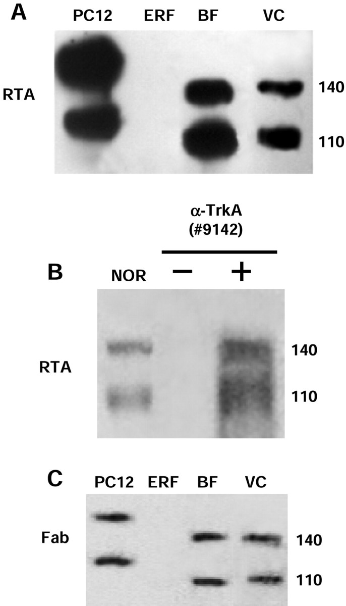
Fig. 1.

RTA immunoblot analysis for detection of the TrkA receptor and control tests for the specificity of the RTA signal.A, Protein extracts were prepared from pheochromocytoma cells (PC12; positive control; 20 μg/lane), epidermal rat fibroblasts (ERF; negative control; 20 μg/lane), basal forebrain (BF; 50 μg/lane), and visual cortex (VC; 200 μg/lane). The 140 kDa band corresponds to the functional form of TrkA receptor, whereas the 110 kDa band is an underglycosylated form of TrkA. B, Western blot of protein extracts (1 mg) from visual cortex of adult rats, immunoprecipitated with the anti-TrkA antibody (#9142; New England Biolabs) and immunoblotted with the RTA antibody. The immunoprecipitated sample (+ α-TrkA) showed a signal similar to the non-immunoprecipitated one [normal (NOR)]. No signal was detected by omitting anti-TrkA (#9142) from the immunoprecipitation step (− α-TrkA). C, The Fab fragment of the RTA antibody was used as the probe. The signal obtained in all positive samples was similar to that obtained using the RTA antibody (for comparison, see A).
