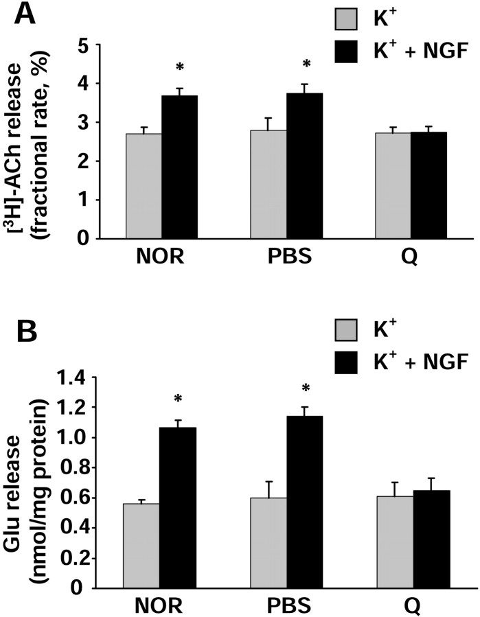
Fig. 5.
Analysis of neurotransmitter release in synaptosomes isolated from untreated [normal (NOR)], PBS-injected (PBS), or quisqualic acid-injected (Q) P30 rat visual cortex. NGF was used at a concentration of 100 ng/ml; KCl was 15 mm. Data are expressed as the mean ± SEM values (n = 12 experiments run in triplicate; evoked release fraction minus averaged basal release fractions). Gray columns represent experiments in which the 90 sec K+-depolarizing stimulus was given alone.Black columns represent experiments in which the K+-depolarizing stimulus was given in the presence of NGF. For each animal group, the values obtained in the presence of NGF have been compared with those obtained in the absence of NGF. Statistical calculations were performed using Student's two-tailedt test; *p < 0.01.A, [3H]Acetylcholine release is expressed as fractional rate (percentage; amount of radioactivity in a fraction divided by the remaining content). Note that the NGF-potentiating effect is almost completely abolished in quisqualic acid-injected animals. B, Endogenous glutamate release is expressed as nanomoles present in the superfusate fraction per milligram of protein of the synaptosomal preparation. The spontaneous basal release did not vary between groups (0.226 ± 0.008 nmol/mg protein). Note the abolition of the NGF effect in BFCN-lesioned animals. For both neurotransmitters, no statistical difference between different groups was detected for the values obtained in the presence of K+ alone (gray columns). NGF had no effect on the spontaneous basal release of both acetylcholine and glutamate in the absence of the K+-depolarizing stimulus (data not shown) (Sala et al., 1998).

