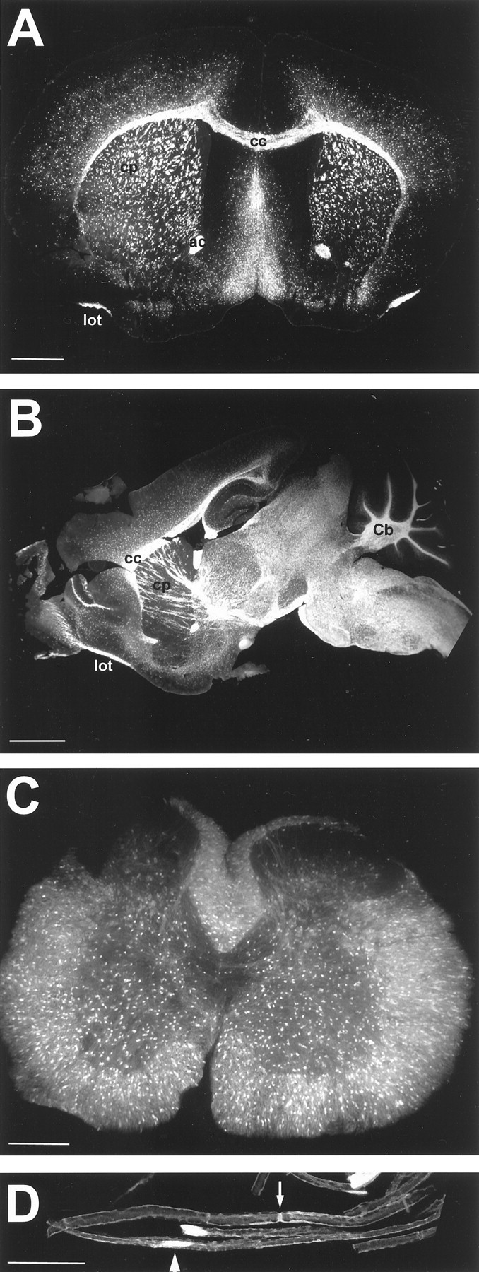
Fig. 1.

Overview of EGFP expression in postnatal nervous system. A, Coronal section of P21 brain (EGFP10) showing vivid fluorescence in white matter tracts, including corpus callosum (cc), caudate putamen (cp), anterior commissure (ac), and the lateral olfactory tract (lot). B, Sagittal section of P21 brain (EGFP5) demonstrating strongest fluorescence in the brainstem and spinal cord, as well as white matter tracts of the cerebellum (Cb). C, Cross section of P30 spinal cord (EGFP3) showing EGFP- positive cell bodies in both gray and white matter.D, Teased fibers of 6 month sciatic nerve (EGFP10) showing fluorescence in cell bodies (arrowhead) and the paranode (arrow). Scale bars: A, 1000 μm; B, 2000 μm; C, 500 μm;D, 25 μm.
