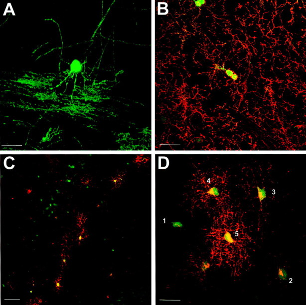
Fig. 3.
Transgene expression may be detected at all stages of the oligodendrocyte lineage. A, EGFP-stained myelinating oligodendrocyte in P21 brain (EGFP5). Note the many parallel processes of myelinated axons. B, NG2 immunostaining of P22 cortex (EGFP10) showing oligodendrocyte progenitor cells. Some NG2-positive cells (Texas Red) are clearly expressing the transgene and are often found in closely apposed pairs or doublets. C, PLP/DM20 (Texas Red) immunostaining of P1 subcortical white matter (EGFP10) showing a clear band of premyelinating oligodendrocytes only in the developing subcortical white matter. D, PLP/DM20 (Texas Red) immunostaining of P1 corpus callosum (EGFP10). Cells in an apparent progression of differentiated states are numbered 1–5, as discussed in Results. Scale bars: A, B,D, 25 μm; C, 50 μm.

