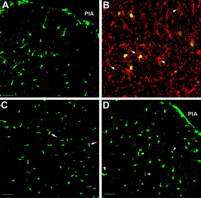
Fig. 5.
Radial migration through the gray matter is complete by P6. A, Many EGFP-positive cells in the outer cortex of P1 brain appear to have a migratory morphology, with some exhibiting a unipolar morphology (EGFP10). B, NG2 immunostaining of P1 outer cortex (EGFP10) showing that the EGFP-positive cells in P1 cortex also express NG2 (Texas Red).Arrowheads indicate nongreen NG2-positive cells.C, By P4, the cells in the outer cortex are beginning to show a few doublets, suggestive of proliferation (seearrows), but some still retain the unipolar shape (EGFP10). D, At P6, the cells populating the outer cortex have lost the unipolar leading process and are often found in pairs (arrowheads; EGFP10). All sections are oriented comparably, with the pial surface at the top right in the image (PIA). Scale bars: A,C, D, 50 μm; B, 25 μm.

