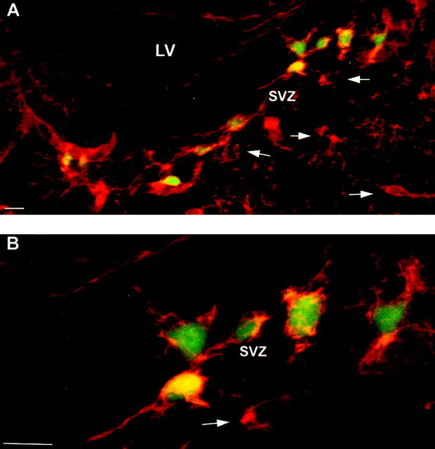
Fig. 7.
EGFP+/NG2+cells are present in the subventricular zone at P1. P1 tissue (EGFP10) was stained for NG2 proteoglycan and imaged for NG2 (Texas Red) and EGFP. A, Image of general SVZ area. LV , Lateral ventricle. B, Higher magnification of cells near the SVZ. Arrows highlight EGFP−/NG2+ cells in the SVZ. Scale bars, 10 μm.

