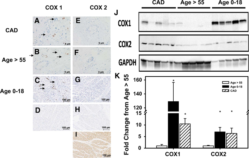
Fig. 2.

Cyclooxygenase expression in human atrial sections. Representative image of IHC in atrial sections for COX1 (a-d) and COX2 (e-i) and Western blot quantification of COX1 and 2 in left ventricle tissue (j, k). COX1 expression was significantly increased in vessels from atrial sections (IHC) and whole left ventricle (WB) tissue from pediatric subjects and patients with CAD compared to adult subjects without CAD. COX2 expression is elevated in vessels from atrial sections (IHC) and whole left ventricle (WB) of CAD or children’s tissue vs. non-CAD controls but not in microvessels of subjects with CAD. d, h Control sections without primary antibody staining. Positive control (i) for COX2 is derived from subjects with autoimmune disease *P < 0.05 two-way t test with Tukey’s post hoc analysis, IHC, N = 4; WB, N = 5
