Fig. 1.
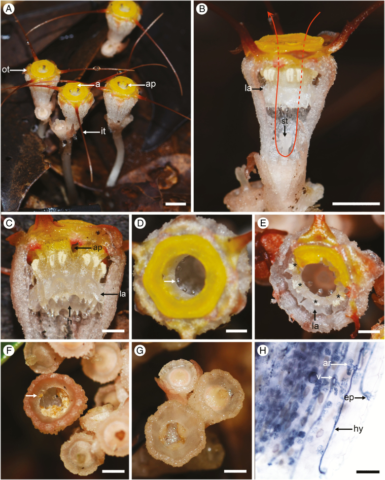
Thismia tentaculata. (A) Several flowering individuals. (B) Dissected flower with proximal part of the perianth tube removed. The unidirectional movement of fungus gnat pollinators is shown by the red arrow (dotted line showing the path behind the annulus and stamens). (C) Dissected flower, showing the staminal ring, with apertures between stamen filaments. (D) Top view of the perianth tube, showing bright yellow annulus and the small droplet of exudate (arrowed) forming at the tip of each pendent hair. (E) The perianth tube, partially destroyed by an insect, showing the lateral appendages; stamen positions are marked by asterisks. (F) A fruit-cup with tiny seeds (arrow shows seed deposited on rim of the fruit-cup due to rain-splash). (G) An empty fruit-cup after rain. (H) Mycorrhizal fungal hyphae in root cortical cells of T. tentaculata. Abbreviations: a, annulus; ap, aperture; ar, arbuscule; ep, entry point; h, hair; hy, hyphae; it, inner tepal; la, lateral appendage; ot, outer tepal; st, stigma; v, vesicle. Photographs by X. Guo. Scale bars: (A, B) = 5 mm; (C–G) = 2 mm; (H) = 100 µm.

