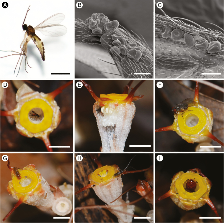
Fig. 3.
Floral visitors to Thismia tentaculata. (A) Fungus gnat (Corynoptera sp.). (B, C) Scanning electron micrographs of pollen grains of T. tentaculata attached to the leg and wing of a Corynoptera fungus gnat. (D) A Corynoptera fungus gnat constrained within the perianth tube. (E) A fungus gnat escaping from the floral chamber via small apertures between the staminal filaments. (F) House fly. (G) Fruit fly. (H) Springtail. (I) Nitidulid beetle. Photographs by X. Guo. Scale bars: (A) = 0.5 mm; (B, C) = 20 µm; (D–I) = 5 mm.

