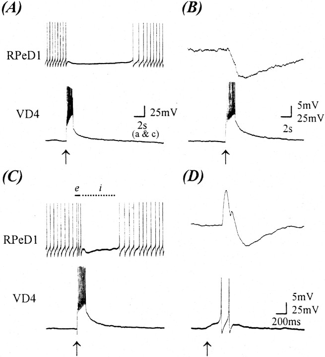
Fig. 1.
CM promotes the formation of an excitatory synaptic component from VD4 to RPeD1 at a normally inhibitory synapse. After 18 hr of cell pairing, simultaneous intracellular recordings from VD4 and RPeD1 somata revealed an inhibitory synapse in DM (A, B), whereas in 100% CM, an excitatory synaptic component (biphasic synapse) was detected (C, D).A, Specifically, in DM, a burst of action potentials in VD4 inhibited spontaneous action potentials in RPeD1. Similarly, action potentials in VD4 produced a compound IPSP in RPeD1 [B; membrane potential (VR), −55 mV]. In contrast, when this synapse was reconstructed in 100% CM, a burst of action potentials in VD4 initially enhanced the rate of spontaneous activity (C, solid line,e) in RPeD1, followed by an inhibition of the firing of action potentials (C, dotted line,i). Likewise, a burst of action potentials in VD4 produced an initial EPSP followed by an IPSP in RPeD1 (D; VR, −55 mV).Arrows indicate the injection of depolarizing current.

