
Fig. 2.
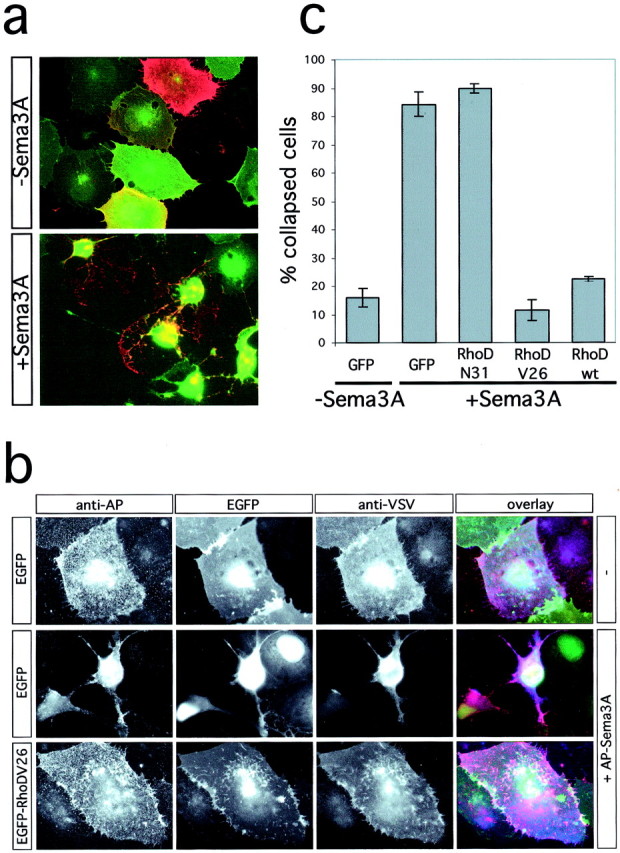
RhoD blocks Sema3A-induced collapse of COS-7 cells. a, COS-7 cells were transfected with the expression vectors pBK-VSV-PlexinA1, pBK-HA-Nrp-1, and pEGFP and incubated with medium (−Sema3A) or with medium containing 0.4 nm AP-Sema3A (+Sema3A) for 1 hr at 37°C. Cells were fixed and processed for immunofluorescence without permeabilization. The expression of recombinant proteins was visualized by EGFP-fluorescence or indirect immunofluorescence using an anti-VSV antibody. b, COS-7 cells were transfected with the expression vectors pBK-VSV-PlexinA1, pBK-HA-Nrp-1, and pEGFP or pEGFP-RhoDV26 as indicated and incubated with medium (−) or with medium containing 0.4 nm AP-Sema3A (+AP-Sema3A) for 1 hr at 37°C. Bound AP-Sema3A was revealed using an anti-alkaline phosphatase antibody (red). The expression of recombinant proteins was visualized by EGFP-fluorescence (green) or indirect immunofluorescence using an anti-VSV antibody (blue). c, The number of cells collapsed in response to Sema3A was determined according to published criteria (Takahashi et al., 1999). The percentage of collapsed cells (gray bars) is displayed (n = 4; 200–300 cells counted per experiment). wt, Wild type.