
Fig. 1.
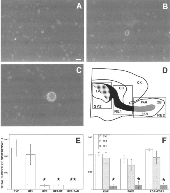
Undifferentiated cells isolated from the RE of adult mice proliferate in response to GFs. The forebrain of adult mice was dissected out, and three different regions were isolated (D): SVZ, subventricular zone;RE1, rostral extension in olfactory peduncle;RE2, rostral extension in the OB. SVZ–RE tissue is indicated in black (the dotted area shows the ventricular wall); RE tissue of the OB obtained by microdissection (not including the surrounding parenchyma) is indicated ingray. LV, Lateral ventricle;CX, cortex; CC, corpus callosum;PAR, parenchyma. Cells were cultured in the presence of EGF, FGF-2, or both; the number of spheres formed in each well was counted after 7–12 DIV. A, Hypertrophic cell from RE2 after 4 DIV proliferates and gives rise after 8 DIV to a small cluster of proliferating cells (B). After 12 DIV a primary sphere is formed (C). E,Spheres were generated from cells isolated from all three regions, but significantly fewer were obtained from RE2-derived cultures compared with those derived from SVZ and RE1. Microdissection and separation of the RE2 region into RE tissue (RE2/RE) and surrounding parenchyma (RE2/PAR), followed by culturing, showed that only cells dissociated from RE2/RE gave rise to spheres.F, Use of EGF, FGF-2, or both generated closely similar numbers of spheres in each of the three regions. Data are means ± SD of four independent experiments in triplicate. Tissue from two mice was pooled in each experiment. Scale bar, 20 μm. *Significantly different from SVZ and RE1; **significantly different from RE2/RE; Student's t test, p < 0.05.