
Fig. 2.
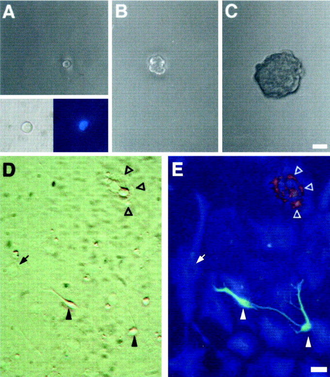
Cells isolated from the RE of adult mice are multipotent. Primary RE-generated spheres were dissociated, single cells were transferred to individual wells by micromanipulation (1 cell per well) in growth medium, and followed by time-lapse microphotography. Virtually all of the cells that were classified as single cells by visual inspection in our clonal assays were indeed single cells, as confirmed by the detection of a single nucleus by DAPI staining (A, inset). The cell shown in A(2 hr after plating; derived from an RE2-primary sphere) proliferated (B; 7 d) and gave rise after 20 d to a spherical clone of cells (primary clonal sphere; C). Primary clonal spheres were subcloned to generate secondary clonal spheres, which were pooled and serially passaged to generate a clonal cell line. After differentiation by removal of GFs, the progeny of RE-derived clonal cell lines included neuronal, astroglial, and oligodendroglial cells. D, E, Phase-contrast and fluorescence micrographs of differentiated cultures from the RE2-derived clonal cell line A7.16. Triple-labeling immunofluorescence revealed the simultaneous presence of neurons (Tuj1,green; filled arrowheads), astrocytes (GFAP, blue; arrows), and oligodendrocytes (O4, red; open arrowheads) within the same culture. Scale bars:A–C, 30 μm; D, E, 20 μm.