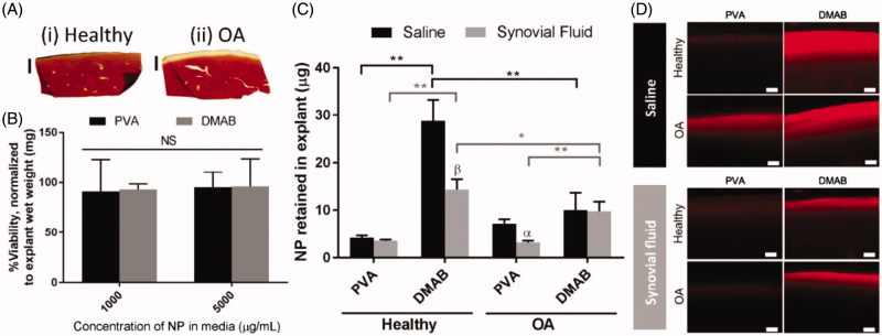
Figure 2.
Ex vivo cartilage retention studies for PLGA NPs, indicating the interaction between NPs and cartilage. (A) Histological sections for (i) health cartilage and (ii) enzymatically digested OA cartilage. (B) Cytotoxicity of PLGA NPs to the cartilage (n = 6). (C) Quantified analysis of NPs’ retained in explant cartilage after incubation (n = 5). (D) Cross section of health and OA sample after incubated with NPs observed with a fluorescence microscopy (Scale bar = 100 µm). α, p < 0.05, β, p < .01 representing the significance between saline and synovial fluid treatments. Reprinted with permission from Brown et al. (2019).

