
Fig. 1.
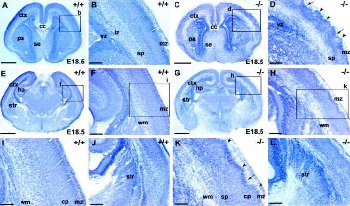
Nissl staining reveals grossly normal development of the telencephalon and diencephalon inSnap25 null mutant mice. Only mild abnormalities in the regularity of the cortical plate were observed in the null mutant. Coronal sections (60 μm) were cut and stained with cresyl violet to reveal major brain structures. The general morphology, cellular distribution, and lamination patterns of WT, heterozygous, and null mutant littermates were compared. A, E, Coronal sections at different rostrocaudal levels of an E18.5 WT brain (+/+). B, F, Higher-magnification views of the boxed areas labeled b andf in A and E, respectively, to demonstrate the normal lamination of the cortex (ctx) at the two levels. I, Detail of theboxed area (i) inF. J, Part of a coronal section from another E18.5 WT brain, showing the patterning of cells of the primordial corpus striatum (str) created by axon bundles of the primitive internal capsule. C, G, Coronal sections at different rostrocaudal levels in theSnap25 null mutant mouse (−/−) at E18.5.D, H, High magnification of the areasboxed in C and G, respectively. Note the abnormal lamination of the cortical plate, particularly in the upper layers and marginal zone (mz), with distinct peaks (arrowheads) and troughs (arrows) along the upper margin of layer 2.K, Detail of the box labeled kin H. Note the abnormal undulation of the upper layers of the cortical plate (arrowheads andarrows). L, View of the cellular patterning created by axonal bundles in the striatum in the null mutant, comparable with that seen in the WT (J). Scale bars: A, C, E,G, 800 μm; B, D,F, J, H, L, 200 μm; I, K, 100 μm.cp, Cortical plate; hp, hippocampus;cc, corpus callosum; se, septal eminence;pa, pallidum; vz, ventricular zone;iz, intermediate zone; sp, subplate;wm, white matter.