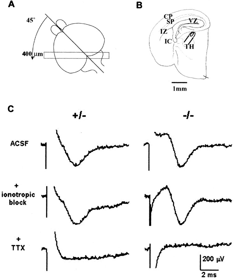
Fig. 8.
Electrophysiological experiments using a whole forebrain slice preparation at E18.5 (Higashi et al., 2002) revealed that thalamic axons conduct APs, but there is no obvious synaptic transmission onto neurons of the cortical plate in WT, HT, or KO brains. A, Diagram to illustrate the plane of vertical section (45° to both coronal and sagittal planes) at which brains were cut at 400 μm to produce slices containing the VB complex of the thalamus, the putative somatosensory cortex, and the entire fiber pathway in between. B, Camera lucida tracing of a thalamocortical slice preparation showing the position of the stimulating electrode in the thalamus (TH).CP, Cortical plate; SP, subplate;IZ, intermediate zone; VZ, ventricular zone; IC, internal capsule. C, Extracellular recordings after thalamic stimulation in slices from HT (+/−, left column) and KO (−/−, right column) brains. In both genotypes, stimulation of the VB produced an initial artifact (the polarity of which depended on the stimulating polarity), followed by a negative-going field potential with a peak at ∼4 msec. This peak was not eliminated by applying ionotropic blockers (40 μm CNQX, 10 μmMK801, 20 μm bicuculline) in the bath. The field potential was eliminated, however, by the subsequent application of 600 nm TTX. The responses are indistinguishable except for a difference in the form of the stimulus artifact.

