
Fig. 3.
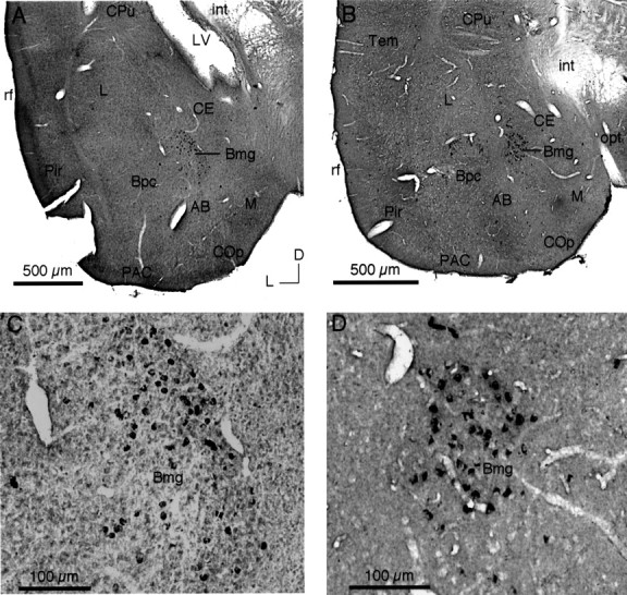
Retrograde labeling in Bmg after tracer deposits in the IC. A, C, Lower-power (A) and higher-power (C) views of retrograde labeling in the ipsilateral Bmg of the mustached bat after the CTb deposit illustrated in Figure2B. B, D, Lower-power (B) and higher-power (D) views of retrograde labeling in the ipsilateral Bmg of the pallid bat after the CTb deposit illustrated in Figure 2D. Retrograde neuronal labeling is restricted to the Bmg. Examples of artifactual label are evident in C (far left) and D (top). Protocol was as follows (plan apochromat): A, B, NA 0.08; C, D, NA 0.40.