
Fig. 4.
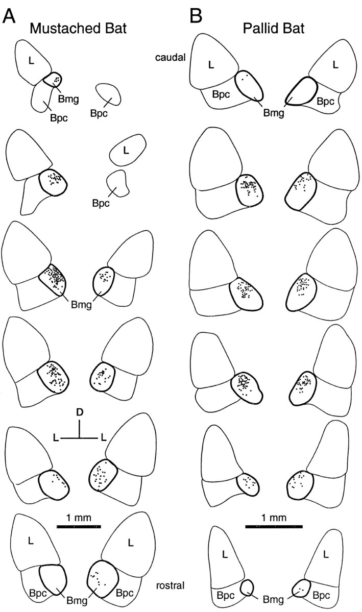
Distribution of retrograde label in the amygdala after tracer deposits in the IC. A, Labeling in a mustached bat after tracer deposit shown in Figure2B. B, Labeling in a pallid bat after tracer deposit shown in Figure 2D. Sections are arranged from caudal (top) to rostral (bottom), and the labeling on the side ipsilateral to the deposit is presented on the left. In both experiments, labeling was bilateral with an ipsilateral predominance. Outline of the Bmg is in bold.