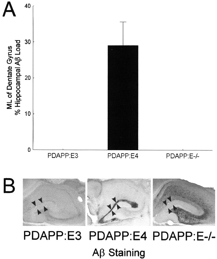
Fig. 1.
A, Almost one-third of the total hippocampal Aβ load was contained in the ML of the dentate gyrus in PDAPP:E4 mice. Localization of Aβ deposition in the ML is associated with the formation of fibrillar amyloid. In contrast, no ML Aβ-IR deposits were found in PDAPP:E3 or PDAPP:E−/− mice 3 months after TBI. B, Photomicrographs show Aβ staining in the hippocampus (arrowheads delineate the borders of the ML).

